
Six points that prevent most MCH mistakes
- MCH is calculated from haemoglobin and red-cell count. It is not independently measured, and it is not an iron level or oxygen-saturation test.See reference 1,See reference 4
- Use the unit and interval printed on your report. Adult examples around 27–32 pg are not a universal optimal range.See reference 1,See reference 2,See reference 9
- MCH is haemoglobin mass per cell; MCHC is concentration inside red-cell volume; MCV is cell volume. They answer different questions.See reference 2,See reference 4,See reference 7
- Low MCH does not prove iron deficiency. Thalassaemia, other haemoglobin disorders, sideroblastic anaemia and some chronic-disease patterns overlap.See reference 5,See reference 6,See reference 11
- High MCH often reflects larger cells, not dangerously concentrated cells. B12/folate, alcohol/liver, thyroid, reticulocyte, pregnancy, medicine and marrow contexts differ.See reference 7,See reference 8
- Do not start iron, B12 or folate, or stop a medicine, from MCH alone. Active bleeding or severe symptoms need care based on the clinical situation, not the MCH flag.See reference 5,See reference 6,See reference 12,See reference 13,See reference 14
What MCH measures—and how the laboratory calculates it
MCH summarizes the average haemoglobin mass in a circulating red cell. With haemoglobin in g/dL and RBC count in millions/µL, the formula is MCH (pg) = haemoglobin × 10 ÷ RBC. With haemoglobin in g/L and RBC in 10^12/L, dividing the two numeric values directly gives pg.See reference 1,See reference 4
Because MCH is derived, it inherits error from either input. A falsely low RBC count can push MCH up; a falsely high measured haemoglobin can do the same. Analyser flags or a biologically implausible CBC pattern may matter more than the displayed index.See reference 4,See reference 10
MCH describes an average, not every cell. It cannot show iron stores, identify a vitamin deficiency, diagnose thalassaemia, grade anaemia, measure oxygen saturation or decide whether a transfusion is needed.See reference 5,See reference 6,See reference 7,See reference 11
Adult examples show why the report interval comes first
- Source or context
- Your reporting laboratory
- Educational MCH example
- The interval printed beside the same result and unit
- Safe interpretation
- Best first comparison because it reflects that method and reference population. It is not a diagnosis, treatment target or emergency threshold.See reference 1,See reference 2,See reference 9
- Source or context
- RCPA adult example
- Educational MCH example
- 27–32 pg
- Safe interpretation
- RCPA states there is method variation and instructs readers to refer to the laboratory.See reference 1
- Source or context
- MedlinePlus adult example
- Educational MCH example
- 27–32 pg/cell
- Safe interpretation
- MedlinePlus says ranges and measurement conventions can differ among laboratories.See reference 2
- Source or context
- Children
- Educational MCH example
- Age-specific examples differ from adults
- Safe interpretation
- Use the child’s age-specific report interval; newborn, infant and child ranges must not be copied from an adult web table.See reference 1,See reference 9
Units, preparation, specimen and retesting
One picogram is 10^-12 grams. ICSH recommends pg for MCH; its international survey found fmol reporting in Denmark and the Netherlands. A clinical laboratory handbook using the haemoglobin-monomer/iron-atom convention gives pg × 0.06206 = fmol, so 27–33 pg is about 1.68–2.05 fmol. Because the elementary entity must be explicit and another report may use a different convention, keep the original unit and interval and do not convert an unlabeled result.See reference 3,See reference 15
MCH is usually calculated from venous EDTA whole blood as part of a CBC/FBC. The CBC itself generally needs no fasting or special preparation; follow separate instructions for other tests collected at the same time.See reference 1,See reference 2
Preserve the exact value, unit or molar entity, interval, date and laboratory flags. There is no universal MCH-specific repeat interval. Unexpected, persistent, changing, implausible or discordant results may need laboratory review or a repeat chosen for the full CBC, symptoms and suspected cause.See reference 1,See reference 9,See reference 10
MCH vs MCV vs MCHC: the difference in one table
- Index
- MCH — usually pg
- What it describes
- Average haemoglobin mass in one red cell
- Common mistake to avoid
- It is not haemoglobin concentration, cell size, iron stores or oxygen saturation.See reference 1,See reference 2,See reference 4
- Index
- MCV — fL
- What it describes
- Average red-cell volume or size
- Common mistake to avoid
- A high or low MCV classifies cell size; it does not give haemoglobin mass per cell.See reference 2,See reference 7
- Index
- MCHC — g/L or g/dL
- What it describes
- Average haemoglobin concentration within red-cell volume
- Common mistake to avoid
- High MCH can occur because cells are larger while MCHC remains normal.See reference 2,See reference 4,See reference 7
- Index
- Haemoglobin/Hb/Hgb — g/L or g/dL
- What it describes
- Haemoglobin concentration in whole blood
- Common mistake to avoid
- Anaemia and clinical severity are not determined from MCH alone.See reference 2,See reference 5,See reference 12
Low MCH: what can reduce haemoglobin mass per cell?
- Possibility
- Iron restriction or iron deficiency
- Why MCH may be low
- Less available iron can limit haemoglobin production and produce hypochromic, often microcytic cells.
- Context that separates possibilities
- Haemoglobin, MCV/RDW, ferritin, transferrin saturation, inflammation and a search for intake, absorption or blood-loss causes. MCH alone is not diagnostic.See reference 5,See reference 6,See reference 12
- Possibility
- Thalassaemia or another haemoglobinopathy
- Why MCH may be low
- Inherited globin-production differences can produce small cells with low MCH.
- Context that separates possibilities
- RBC count and family/ancestry context can suggest a pattern, but iron studies and clinician-directed haemoglobin or genetic analysis are needed; MCH/MCV cannot distinguish the diagnosis.See reference 5,See reference 11
- Possibility
- Chronic inflammation or illness
- Why MCH may be low
- Functional iron restriction and altered red-cell production can lower MCH in some cases.
- Context that separates possibilities
- Inflammatory, kidney and clinical context; ferritin may be affected by inflammation.See reference 5,See reference 6
- Possibility
- Sideroblastic or other less common processes
- Why MCH may be low
- Haem synthesis can be impaired despite iron being present.
- Context that separates possibilities
- Medicine/exposure history, smear and specialist testing when common explanations do not fit.See reference 5,See reference 7
High MCH: usually a large-cell pattern, sometimes an artefact
- Possibility
- Vitamin B12 or folate deficiency
- Why MCH may rise
- Impaired DNA synthesis can produce larger developing red cells that carry more total haemoglobin per cell.
- Context that helps
- MCV/RDW, symptoms, diet/absorption/medicine history, B12/folate testing and smear. MCH itself cannot identify the deficiency.See reference 7,See reference 8
- Possibility
- Alcohol exposure or liver disease
- Why MCH may rise
- Direct marrow and red-cell membrane effects can produce macrocytosis and high MCH.
- Context that helps
- Alcohol history, liver tests, platelet count and the wider CBC; do not infer alcohol use from MCH alone.See reference 7,See reference 8
- Possibility
- Hypothyroidism or reticulocytosis
- Why MCH may rise
- Thyroid disease can accompany macrocytosis; larger immature reticulocytes can raise averages after bleeding or haemolysis.
- Context that helps
- TSH, reticulocytes, bilirubin/LDH/haptoglobin, smear and bleeding context as clinically appropriate.See reference 7,See reference 8
- Possibility
- Medicines or marrow disease
- Why MCH may rise
- Selected antimetabolite, chemotherapy, hydroxycarbamide and antiretroviral medicines alter cell development; marrow disorders are less common alternatives.
- Context that helps
- Medication review, WBC/platelets, smear and clinician assessment. Never stop a prescription from MCH alone.See reference 8
- Possibility
- Calculated-result or specimen artefact
- Why MCH may rise
- Cold agglutinins can falsely lower RBC count; haemolysis, lipaemia, extreme bilirubin or hyperleukocytosis can distort Hb or related indices.
- Context that helps
- An implausible MCH/MCHC, analyser flag, Hb–HCT mismatch or sudden discontinuity should be reviewed by the laboratory rather than corrected at home.See reference 4,See reference 10
Low MCH with normal haemoglobin—and other isolated patterns
A mildly low MCH can appear before haemoglobin crosses an anaemia threshold, with iron-restricted red-cell production, or in an inherited microcytic pattern such as thalassaemia trait. It does not prove any one cause and should be read with MCV, RBC, RDW, ferritin/iron context and trend.See reference 5,See reference 6,See reference 11
Normal MCH does not rule out anaemia, early iron deficiency, B12/folate deficiency, blood loss, kidney/inflammatory disease or mixed small-plus-large cell populations. An average can look ordinary while the distribution is not.See reference 5,See reference 6,See reference 7
An isolated high MCH should first be compared with MCV, MCHC, RBC, haemoglobin and analyser comments. A mathematically high value driven by a very low RBC denominator may need specimen review before a disease explanation.See reference 4,See reference 7,See reference 10
When the sample or analyser can mislead
- Issue
- Cold-reactive red-cell agglutinins
- Possible pattern
- Falsely low RBC/HCT with falsely high MCV, MCH and MCHC
- Safer response
- The laboratory can review flags/smear and use a validated warming or recollection process; the reader should not adjust the value.See reference 10
- Issue
- Lipaemia, extreme bilirubin, hyperleukocytosis or lysis-resistant cells
- Possible pattern
- Falsely high measured haemoglobin can propagate into MCH/MCHC
- Safer response
- Use analyser flags and laboratory procedures; repeat or correct only through the performing laboratory.See reference 10
- Issue
- In-vitro haemolysis
- Possible pattern
- RBC/HCT may fall while MCH/MCHC rise
- Safer response
- A properly collected replacement sample or laboratory comment may be needed; in-vivo haemolysis is a different clinical question.See reference 10
- Issue
- Clot, poor fill, delayed handling or IV-fluid dilution
- Possible pattern
- One or several CBC values may be unreliable or unexpectedly low
- Safer response
- Preserve the analyser comments and recollect under laboratory guidance when advised.See reference 9,See reference 10
Age, pregnancy, treatment and laboratory context
- Context
- Newborns, infants and children
- Why interpretation changes
- MCH distributions change with age, and RCPA examples differ across early life and childhood.
- Safer approach
- Use the age-specific interval on the child’s report and paediatric clinical context.See reference 1,See reference 9
- Context
- Pregnancy
- Why interpretation changes
- Plasma volume, nutrient needs and physiological macrocytosis can change the CBC pattern.
- Safer approach
- Use gestational and local obstetric guidance, symptoms, haemoglobin and the complete report; do not apply one adult web interval.See reference 8
- Context
- Treatment or medicine exposure
- Why interpretation changes
- Iron/B12/folate treatment, transfusion, reticulocyte response and medicines affecting DNA synthesis can change the pattern over time.
- Safer approach
- Record timing and review with the prescriber; do not stop or change treatment from MCH alone.See reference 5,See reference 8,See reference 12
- Context
- Laboratory, analyser or unit change
- Why interpretation changes
- Reference populations, methods and pg/fmol conventions differ.
- Safer approach
- Compare the same laboratory where practical and keep each value with its own unit and interval.See reference 1,See reference 3,See reference 9
Symptoms and haemoglobin matter more than the MCH number
Use urgent local medical care for uncontrolled or major bleeding; vomiting blood; black or bloody stool with weakness; severe or worsening breathlessness; chest pain; fainting; confusion or difficult waking; sudden weakness, speech or vision change; or rapidly worsening paleness or jaundice. Do not wait for a calculator or routine repeat. There is no universal MCH emergency cutoff, and a normal MCH cannot rule out major bleeding, severe anaemia, haemolysis, heart or neurological emergencies.See reference 12,See reference 13,See reference 14
What may change MCH: treat the verified cause
- Pattern
- Low MCH with confirmed iron deficiency
- Cause-directed approach
- Treat the deficiency and investigate why iron is low—such as bleeding, intake or absorption—under appropriate clinical guidance.
- Unsafe shortcut
- Starting iron from MCH alone or ignoring the cause of iron loss.See reference 5,See reference 6,See reference 12
- Pattern
- Low MCH with normal iron studies
- Cause-directed approach
- Consider inherited haemoglobin context and clinician-directed testing when the CBC pattern fits.
- Unsafe shortcut
- Assuming iron deficiency or using a screening formula as a diagnosis.See reference 5,See reference 11
- Pattern
- High MCH with macrocytosis
- Cause-directed approach
- Review B12/folate, alcohol/liver, thyroid, reticulocyte, pregnancy, medicine and marrow context.
- Unsafe shortcut
- Taking folate alone, assuming cancer or stopping a prescription.See reference 7,See reference 8
- Pattern
- Implausible or abrupt MCH change
- Cause-directed approach
- Ask the laboratory to review the sample, analyser flags and source Hb/RBC values before interpreting the change.
- Unsafe shortcut
- Applying a home correction or treating a likely artefact.See reference 4,See reference 10
Common MCH misconceptions
- Claim
- Low MCH proves iron deficiency
- More accurate answer
- No. Iron deficiency is common, but thalassaemia/haemoglobinopathy, sideroblastic and chronic-disease patterns overlap.See reference 5,See reference 6,See reference 11
- Claim
- High MCH proves B12 deficiency
- More accurate answer
- No. High MCH often follows high MCV; folate, alcohol/liver, thyroid, reticulocytes, pregnancy, medicines, marrow disease and artefact are alternatives.See reference 7,See reference 8,See reference 10
- Claim
- MCH and MCHC are the same
- More accurate answer
- No. MCH is mass per cell; MCHC is concentration within red-cell volume.See reference 2,See reference 4,See reference 7
- Claim
- High MCH means each cell is dangerously over-concentrated
- More accurate answer
- Usually not. Larger cells can carry more total haemoglobin while MCHC stays normal.See reference 7
- Claim
- 27–33 pg is universal and optimal
- More accurate answer
- No. Published examples differ, and age, laboratory, analyser, population and reporting unit matter.See reference 1,See reference 2,See reference 3,See reference 9
- Claim
- Abnormal MCH means cancer
- More accurate answer
- No. MCH is nonspecific. Persistent abnormalities with other cytopenias, smear changes or symptoms need assessment without turning one index into a cancer test.See reference 7,See reference 8
A safe sequence after an MCH result
- Confirm the unrounded value, exact pg or fmol convention, report limits, date and analyser flags.See reference 1,See reference 2,See reference 3,See reference 9
- Read haemoglobin, RBC, MCV, MCHC, RDW, reticulocytes, WBC, platelets and any smear comment before naming a cause.See reference 2,See reference 5,See reference 7
- Record symptoms, active or recent bleeding, pregnancy, diet/absorption, family context, alcohol exposure, illness, transfusion and medicines.See reference 8,See reference 12,See reference 13
- Use cause-directed tests and a repeat interval chosen for the whole pattern; do not copy a universal panel or timeline.See reference 5,See reference 6,See reference 9
- Use urgent care for major bleeding, chest pain, severe breathlessness, fainting, confusion, neurological change or rapidly worsening paleness/jaundice.See reference 13,See reference 14
What this guide cannot settle
No worldwide MCH interval, optimal longevity target, pregnancy table, child table, unit conversion, analyser correction, biological-variation threshold or repeat schedule applies to every person and laboratory.See reference 1,See reference 3,See reference 9
MCH can help describe an anaemia pattern but cannot identify the cause, severity or urgency alone. Haemoglobin, symptoms, speed of change, other cell counts, reticulocytes, smear, laboratory review and cause-directed tests can change the meaning.See reference 5,See reference 6,See reference 7,See reference 10,See reference 13
Put MCH in the context of the whole result
Upload a laboratory report to LongevityMate to organise MCH beside haemoglobin, RBC count, MCV, MCHC, RDW, reticulocytes, ferritin, B12, folate, thyroid/liver markers and prior results. You receive structured educational context for discussion—not a diagnosis, emergency decision or treatment prescription.
Understand your lab resultsQuestions people ask about MCH
What is MCH on a blood test?
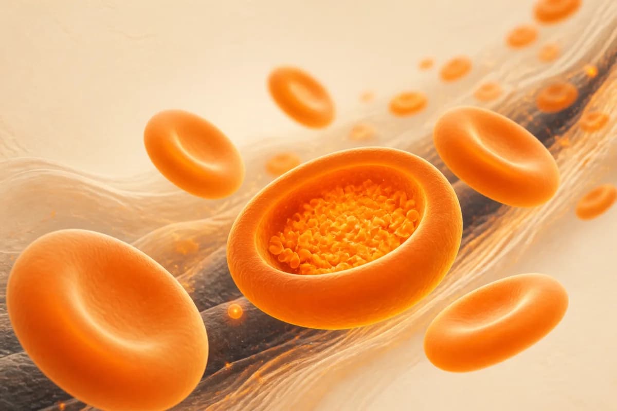
MCH means mean corpuscular haemoglobin or mean cell haemoglobin. It is the calculated average mass of haemoglobin in one red blood cell, usually reported in pg as part of a CBC/FBC.See reference 1,See reference 2,See reference 4
What is a normal MCH range?
Use the interval printed beside your result. RCPA and MedlinePlus both give 27–32 pg as adult educational examples, while stressing laboratory or method variation; children need age-specific intervals.See reference 1,See reference 2,See reference 9
What does low MCH mean?
It means the average red cell contains less haemoglobin mass than the report interval. Iron restriction is one possibility, but thalassaemia/haemoglobinopathy, sideroblastic and some chronic-disease patterns can overlap.See reference 5,See reference 6,See reference 11
Can MCH be low while haemoglobin is normal?
Yes. A low MCH can appear before haemoglobin crosses an anaemia threshold or in an inherited microcytic pattern. Compare MCV, RBC, RDW, ferritin/iron context and trend rather than diagnosing from the isolated flag.See reference 5,See reference 6,See reference 11
Does low MCH always mean iron deficiency?
No. MCH can support an iron-restricted pattern but has limited specificity. Thalassaemia trait cannot be separated from iron deficiency by MCH/MCV alone.See reference 5,See reference 11
What does high MCH mean?
High MCH often follows a high MCV because larger red cells carry more total haemoglobin. B12/folate, alcohol/liver, thyroid, reticulocyte, pregnancy, medicine, marrow and analyser-artifact contexts differ.See reference 7,See reference 8,See reference 10
Is high MCH the same as high MCHC?
No. MCH is haemoglobin mass per cell; MCHC is haemoglobin concentration within red-cell volume. MCH can be high because cells are large while MCHC stays normal.See reference 2,See reference 4,See reference 7
Can high MCH be a laboratory error?
Yes. Because MCH is Hb divided by RBC, cold agglutinins that lower measured RBC or interferences that raise measured Hb can push it up. Analyser flags, MCHC and Hb–HCT consistency help the laboratory recognize an unreliable pattern.See reference 4,See reference 10
Can abnormal MCH mean cancer?
MCH is not a cancer test. Many nutritional, inherited, liver, thyroid, medicine, reticulocyte and analytical explanations exist. Persistent changes with other low cell counts, smear abnormalities or symptoms deserve clinician assessment.See reference 7,See reference 8,See reference 10
Do I need to fast for an MCH test?
Usually not. MCH is part of a CBC/FBC, which generally needs no special preparation, but another test collected at the same time may have separate instructions.See reference 1,See reference 2
Can I convert MCH from pg to fmol?
Only with the laboratory’s explicit molar entity convention. One documented haemoglobin-monomer/iron-atom convention uses 1 pg = 0.06206 fmol, but another convention may differ. Keep the original unit, entity and report interval together; this page’s tool never converts them.See reference 3,See reference 15
When should an abnormal MCH be repeated?
There is no universal MCH-specific timing. Unexpected, persistent, changing, implausible or discordant results may need laboratory review or repeat testing, while active bleeding or severe symptoms need prompt care rather than waiting.See reference 9,See reference 10,See reference 13,See reference 14
Get the weekly longevity brief
The week's best longevity research in plain English. Free, every Tuesday.
References
- 1. Mean cell haemoglobin
Royal College of Pathologists of AustralasiaOfficial guidance
- 2. CBC blood test
MedlinePlus Medical EncyclopediaOfficial guidance
- 3. Recommendation for standardization of haematology reporting units used in the extended blood count
International Council for Standardization in HaematologyGuideline
- 4. Complete Blood Count using HMX: Laboratory Procedure Manual
CDC/NCHS NHANESOfficial guidance
- 5. British Society of Gastroenterology guidelines for the management of iron deficiency anaemia in adults
GutGuideline
- 6. Guideline for the laboratory diagnosis of iron deficiency in adults excluding pregnancy and children
British Society for HaematologyGuideline
- 7. Red Cell Indices
NCBI Bookshelf, Clinical MethodsEvidence review
- 8. Macrocytosis in Adult Patients, A004 v9
Norfolk and Norwich University Hospitals NHS Foundation TrustGuideline
- 9. ICSH guidelines for the evaluation of blood cell analysers
International Council for Standardization in HaematologyGuideline
- 10. Unreliable Automated Complete Blood Count Results: Causes, Recognition, and Resolution
Annals of Laboratory MedicineEvidence review
- 11. Update in Laboratory Diagnosis of Thalassemia
Frontiers in Molecular BiosciencesEvidence review
- 12. Iron-Deficiency Anemia
National Heart, Lung, and Blood InstituteOfficial guidance
- 13. Anemia Symptoms
National Heart, Lung, and Blood InstituteOfficial guidance
- 14. Recognizing medical emergencies
MedlinePlus Medical EncyclopediaOfficial guidance
- 15. Laboratory Service Directory: MCH
BG Kliniken Berlin, Central LaboratoryOfficial guidance
Editorial transparency
- Published by
- LongevityMate Editorial
- Editorial review by
- Lukas Dvorsky, Founder — editorial review
- Published
- Updated
Medical disclaimer
Educational information only, not an anaemia, iron, vitamin B12, folate, thalassaemia, thyroid, liver, haemolysis, marrow or cancer diagnosis; supplement, medicine, transfusion or emergency decision; or a personal target. Use the original laboratory report and qualified clinical care for individual decisions.